As part of the Wallace group at King’s College London, I was investigating label-free optical imaging of single molecules, using Interferometric Scattering (iSCAT) microscopy. Using the fact that all particles can scatter light beams and that this scattered beam can interfere with a reference light beam, we were able to image on our microscope individual metal particles as small as 2nm and individual proteins and polymers as small as 100kg/mol at ultra high speeds (> 10kHz). Going beyond the limitations in space and time resolutions of other types of optical microscopes, we used this technique to visualize key events and reactions in cells and other standard biophysical systems.
Description
Technology
Imaging ever smaller molecules is a constant challenge in research, which lead in the past to the development of new microscopy techniques, such as the electron microscopy techniques or new fluorescence methods to image single molecules. While the former achieves remarkable details, it cannot image a live biological process or track molecular interactions. And while the latter can record diffusion and interactions of single molecules, it relies on the fluorescent probe conjugated with the molecule and is limited by the drawbacks of the probes: price and complexity of the conjugation, photobleaching, limits in time resolution, etc.
The iSCAT microscopy, initiated in the 1990s and developed then mostly through the impulsion of Vahid Sandoghdar’s group, is a microscope technique which allow to track the diffusion and interactions of single molecules without the drawbacks of the fluorescence: the technique can be used directly with any label-free molecule (e.g. nanoparticles, proteins, polymers), and its speed is only limited by the technology of the camera and light source (i.e. up to 1,000,000 FPS with ultra-high-speed cameras).
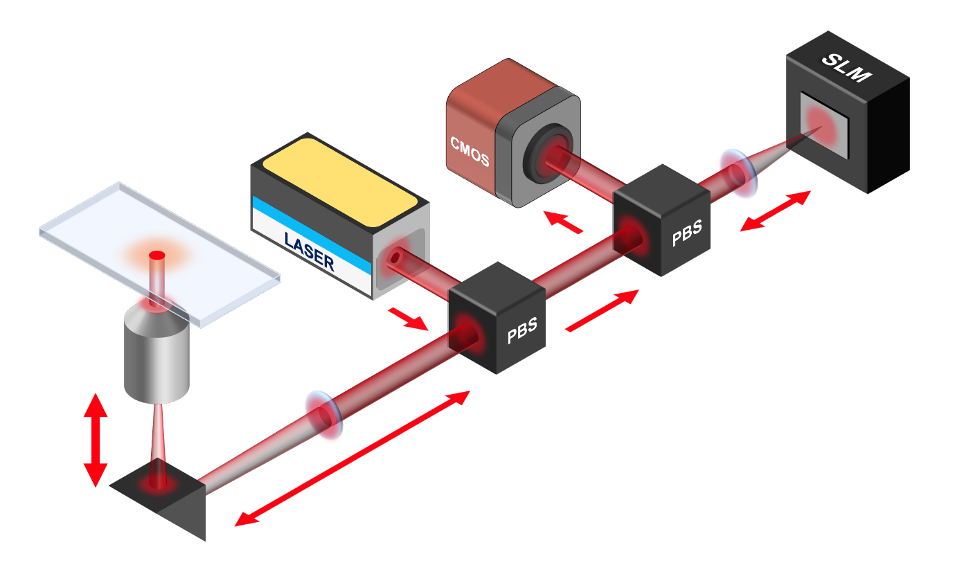
For this project, I have built my own iSCAT microscope, which design can be described simply as it only requires a coherent light source (Laser), a microscope objective, a polarised beam splitter with a wave plate, and the camera for high-speed recording.
Systems
iSCAT microscopy can be applied to any kind of system, as the method does not require any specific property from the object imaged. For this project, we focussed on two topics:
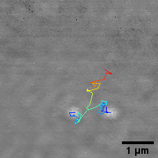
We first investigated in this project the formation of membrane pores in lipid bilayers. With the iSCAT, we can track proteins diffusing on the membrane, and analyse all the interactions leading to the formation of pores and the kinetics associated to these events.
Exploring more artificial soft matter problematics, we also studied polymers with the iSCAT. The microscopy technique allowed us to unveil new insights on the polymer properties and formations.
Supervision & Collaboration(s)
This project was supervised by Prof. Mark I Wallace from the Department of Chemistry of the King’s College London (London, UK). I was myself co-supervising, along with Prof. Wallace, the PhD projects of Christopher L Parperis and Yujie Guo, respectively on membrane pores and polymers imaged in iSCAT.
The whole project was financed through a BBSRC grant with Oxford Nanopore Technologies.
The investigation on polymers were made within a collaboration with Prof. Rachel O’Reilly and Dr. Jeffrey Foster from the University of Birmingham (Birmingham, UK).
Related publication(s)
- Walter et al., Responsive point spread function engineering for interferometric scattering microscopy, submitted to J Phys D 2021