Description
Various possibilites are being investigated for the treatment of cancers. One of these possibilities is to bring inside the tumorous cells a molecule which will induce the cell death, a method called drug delivery. The design of a system for drug delivery requires two parts: the drug, or molecular cargo, which is expected to treat the cell, and the carrier, which has the role of bringing the cargo inside the target cells. Another design consists on encapsulating the drug inside an artificial virus-like capside, to bring the drug inside the cell.
The cell-penetrating peptides (CPP) are a class of oligopeptides which are able to translocate easily through the different types of membranes one can encounter inside living cells, which make them desirable for drug delivery applications. However, cell-penetrating peptides have several drawbacks, like their lack of selectivity toward the target cells, and the fact that their penetrating capacities are disabled when a molecular cargo is attached onto them. In this project, we used cell-penetrating peptides grafted onto elastin-like polypeptides, a diblock copolymer synthetised by the Chilkoti group (Duke University, USA) which has the ability to self-assemble into micelles above a defined temperature.
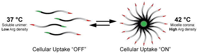
As a unimer, the Elastin-like polypeptide part of the CPP-ELP plays the same role as a molecular cargo, and prevents the CPP to translocate in the cell. Upon self-assembly the CPP recovers its penetrating capacity. We suggest that the increase of the local amount of charges, and specifically of arginine groups, is the key to trigger the penetration of the CPP. This penetration controled by the temperature allows us to induce a selectivity, which the CPP formerly lacked. Indeed, tumourous cells are known to have poor heat exchanges, meaning that a local heating and cooling of a tissue will leave the tumourous cells at a higher temperature than the rest of the cells.
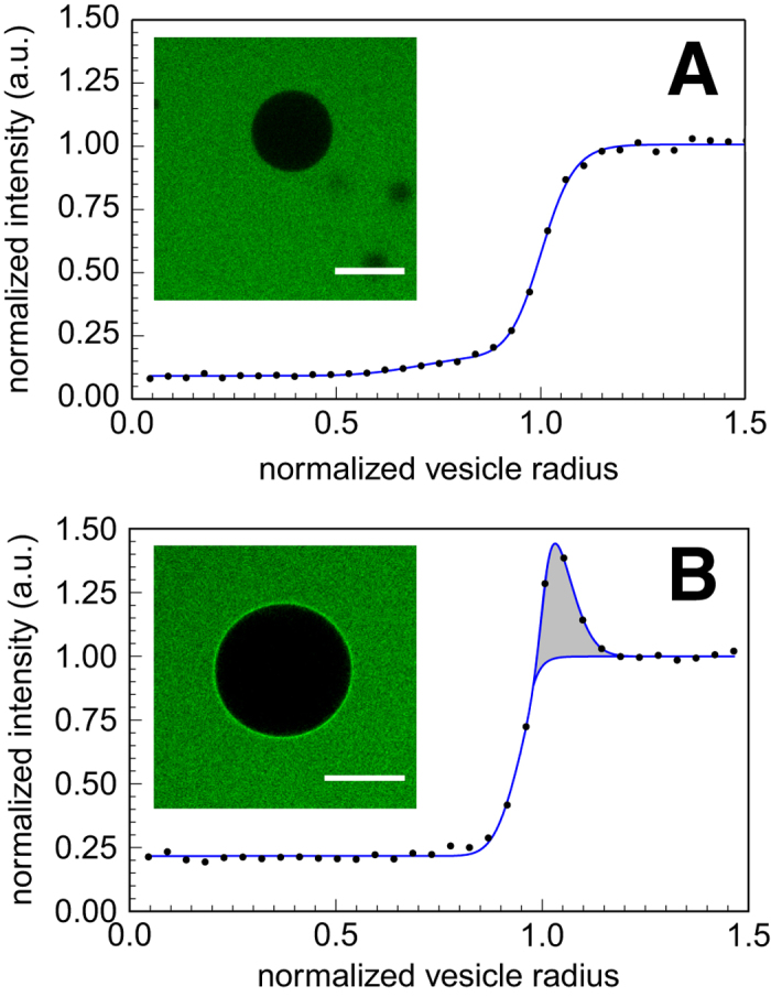
When brought in contact with model lipid membranes such as giant unilamellar vesicles, a spherical self-assembled artificial construction of lipids, we have shown that the CPP-ELP are unable to translocate through the membrane. However, unlike their unimer counterparts, the micelles of CPP-ELP adsorb on the lipid membrane. Since it has been shown that the single CPP sequences are capable of penetrating the model lipid membranes, we hypothesized that the penetration of the CPP is a two step process:
- The CPP reaches the cell membrane and adsorbs onto it, bringing its cargo with it if one is attached.
- The CPP is internalized by the cell, using both passive and active processes (endocytosis, etc.). Due to their size, CPPs grafted onto a molecular cargo can not translocate through the membrane using passive diffusion, and require active processes, which explain the differences in result between cell and vesicles.
Our project then focused on the adsorption of the CPP onto the lipid membrane, and we are using confocal laser scanning microscopy (CLSM) to measure and quantify the number of molecules adsorbed on the membrane. We published this method in 2017. We developed and extended the method to use the confocal microscope to perform thermodynamic measurements on the adsorption of molecules.
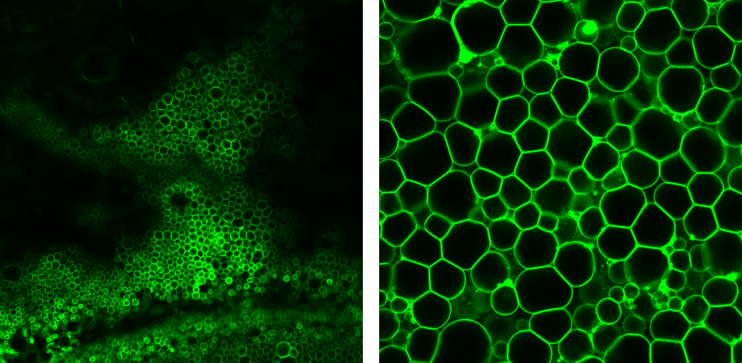
We are also interested in extanding the various protocoles for vesicles formation. We developed a new way to easily obtain giant unilamellar vesicles in a saline solution (up to 150 mM of NaCl) and various buffers, based on the PVA-gel assisted formation previously developed by the team. This method allows us to produce a large number of big sized giant unilamellar vesicles, despite the presence of an important concentration of salt, and we are therefore capable to work easily in typical physiological conditions.
We used this method to investigate the interactions between YOYO-labelled DNA and various types of giant unilamellar vesicles, designed with zwitterionic lipids only (DOPC), negatively charged lipids only (DOPG), and positively charged lipids only (DOTAP). We further extended the protocole to encapsulate biomaterials in vesicles.
Supervision & Collaboration(s)
This project was supervised by Drs. Carlos M Marques, Pierre Muller, Andre Schroeder and Tatiana Schmatko from the Institut Charles Sadron (CNRS, Universite de Strasbourg, FR).
The synthesis of the CPP-ELPs was performed by Prof. Ashutosh Chilkoti and Dr. Sarah MacEwan from Duke University, USA) as part of our collaboration.